
Figure 9.
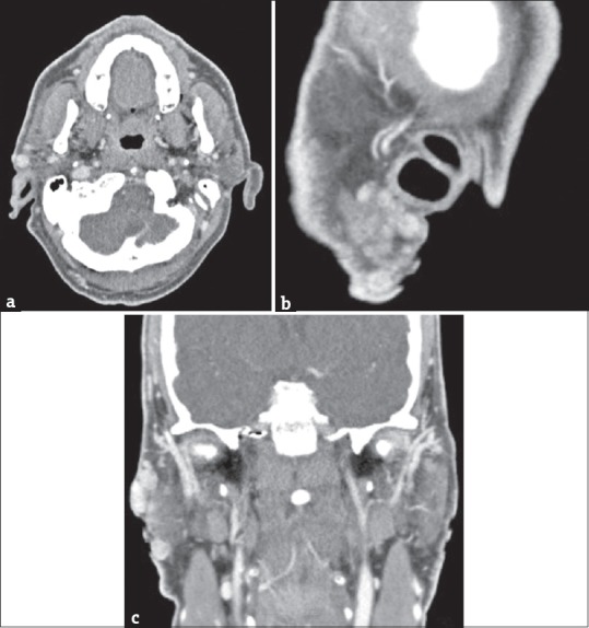
(a) A 58-year-old male postsurgical recurrences in the site of previous right parotidectomy. Axial contrast-enhanced computed tomography scan and multiplanar reformatted images, obtained along sagittal. (b) A 58-year-old male sagittal plane. (c) A 58-year-old male coronal planes show multiple parotid lesions in different locations.