Fig. 1.
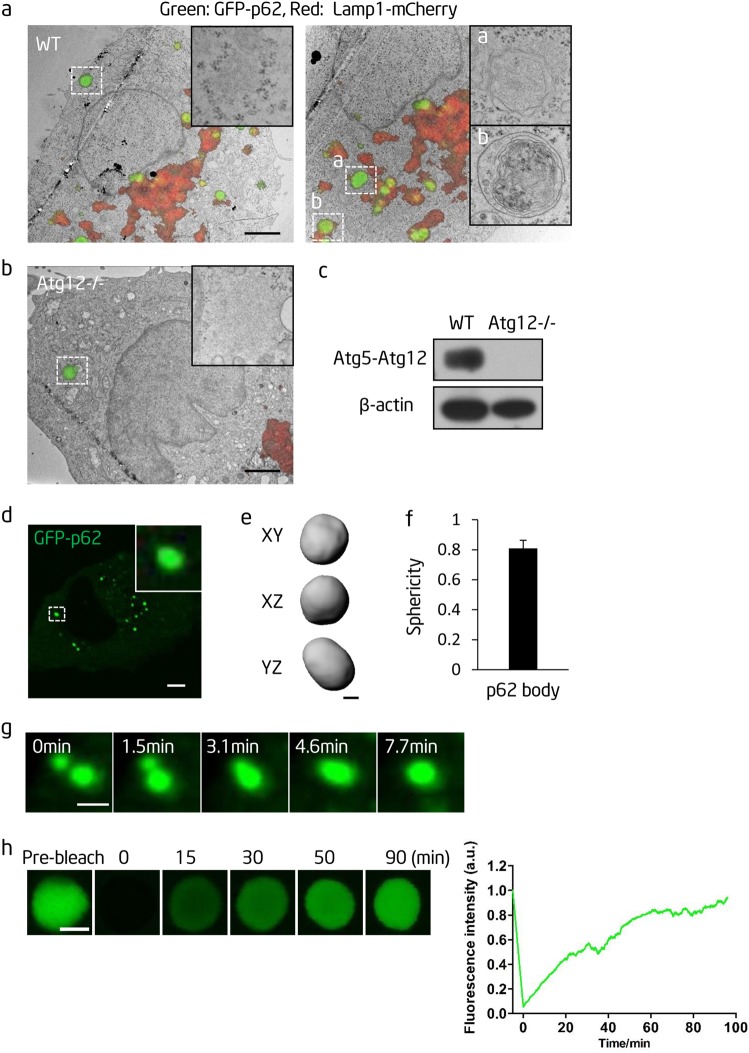
p62 forms liquid droplets in vivo. a Correlative light-electron microscopy (CL-EM) of NRK cells transiently transfected with GFP-p62 and Lamp1-mCherry constructs. Scale bar, 2 µm. The insert in the left panel shows a p62 body. The inserts in the right panel show p62 bodies contained within an autophagosome (upper) and an autolysosome (lower). b CL-EM of Atg12−/− cells transiently transfected with GFP-p62 and Lamp1-mCherry constructs. Scale bar, 2 µm. The insert shows a p62 body. c Western blot analysis of wild-type and Atg12−/− cells with the indicated antibodies. d GFP-labeled p62 forms p62 bodies in Atg12−/− NRK cells. An enlargement of the boxed p62 body is shown in the insert. Scale bar, 5 µm. e Rendered 3D shapes of a p62 body. Cells were fixed with 4% PFA. The panels show the XY, XZ, and YZ planes. Scale bar, 1 µm. f A plot showing the sphericity of p62 bodies (n = 44). Error bar represents SD. g Fusion of p62 bodies. Scale bar, 1 µm. h Left panels: fluorescence intensity recovery of a p62 body after photobleaching. Scale bar, 2 µm. Right panel: quantification of fluorescence intensity recovery of a photobleached p62 body

