Fig. 4.
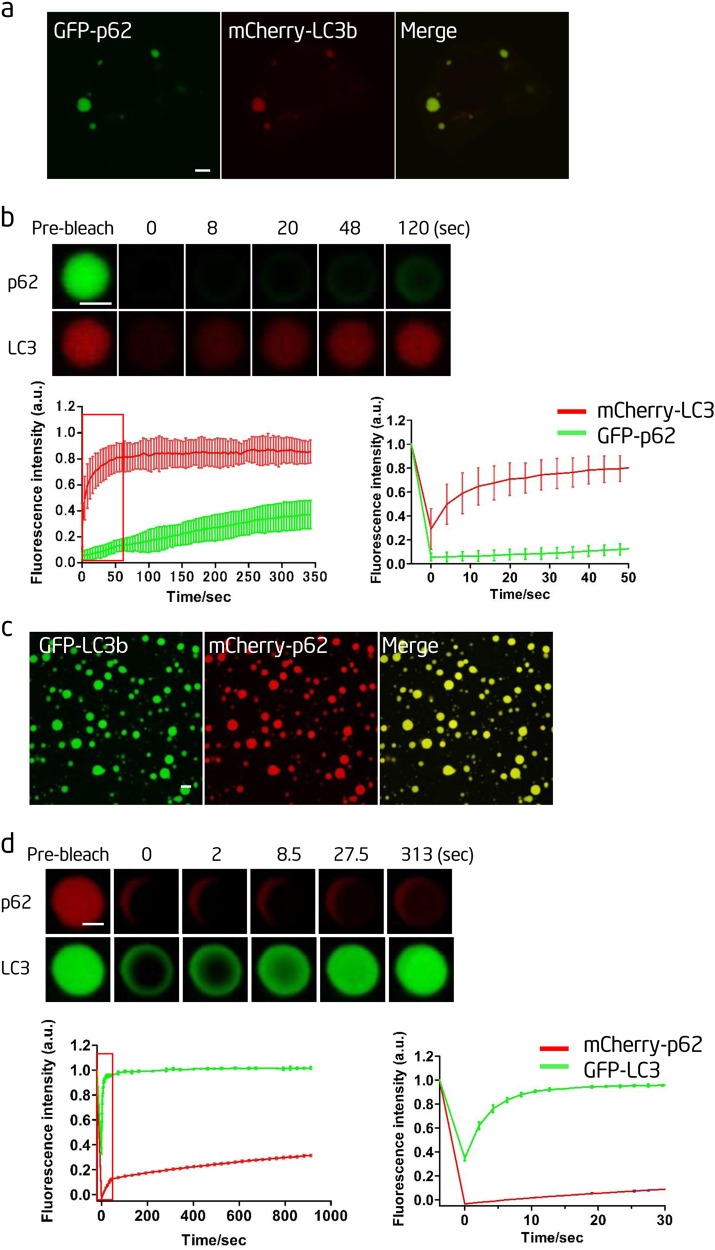
LC3 is recruited into p62 bodies. a Atg12−/− NRK cells were transfected with mCherry-LC3b and GFP-p62 and observed by confocal microscopy. Scale bar, 5 µm. b Upper panels: fluorescence intensity recovery of GFP-p62 and mCherry-LC3b in a bleached p62 body. Scale bar, 2 µm. Bottom left panel: quantification of fluorescence intensity recovery of GFP-p62 and mCherry-LC3b in the bleached p62 body. Bottom right panel: enlargement of the boxed area in the left panel (n=5). c In vitro phase separation assay of mCherry-p62 with linear Ubx8. GFP-LC3b was added after the phase separation. mCherry-p62, 20 µM; Ubx8: 5 µM; GFP-LC3b, 10 µM. Scale bar, 10 µm. d Upper panels: fluorescence intensity recovery of GFP-LC3b and mCherry-p62 in an in vitro-formed mCherry-p62 droplet from c after photobleaching. Scale bar, 2 µm. Bottom left panel: quantification of fluorescence intensity recovery of GFP-LC3b and mCherry-p62 in the bleached p62 droplet. Bottom right panel: enlargement of the boxed area in the left panel (n=3)

