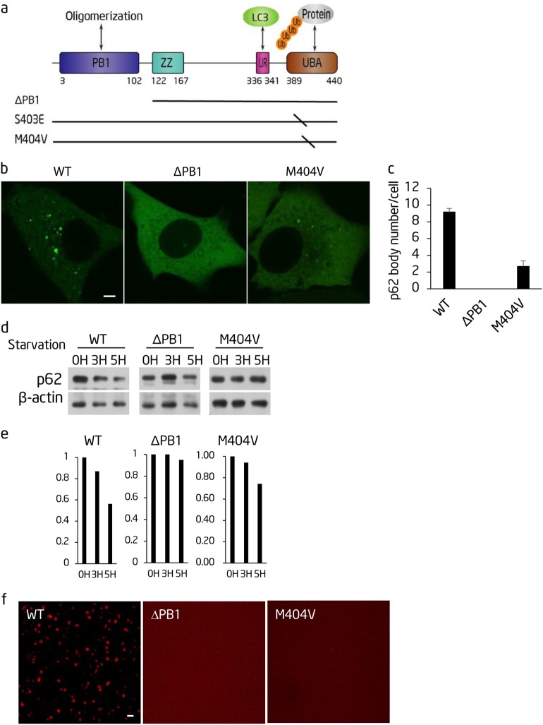
Fig. 5.
The PB1 domain and UBA domain of p62 are required for polyubiquitin chain-induced phase separation and autophagic degradation of p62. a Schematic diagram of the p62 protein domain structure. The location of p62 mutations is shown underneath. b The indicated GFP-p62 constructs were expressed in Atg12−/− cells and observed by confocal microscopy. Scale bar, 5 µm. c. Cells from b were quantified for the number of p62 bodies. >45 cells from b were assessed blind and quantified. Error bars indicate SD (n = 3). d NRK cells transiently expressing the indicated GFP-p62 constructs were starved for the indicated time. Cell lysates were analyzed by western blot with the indicated antibodies. e Quantification of the indicated western blots in d. f The indicated mutated recombinant mCherry-p62 proteins were mixed with linear Ubx8 and the reaction was visualized by confocal microscopy. mCherry-p62, 5 µM; linear Ubx8, 2.5 µM. Scale bar, 5 µm

