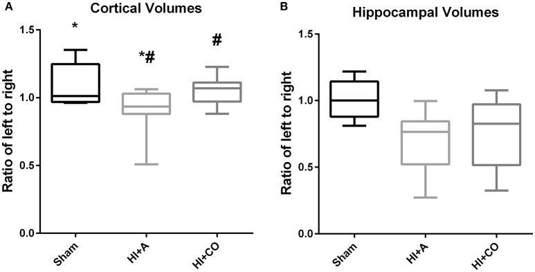
Figure 3.
Cortical and hippocampal volumes. At 12 days of age (5 days post injury), the brain region volumes are represented as the ratio of the left (injured)/right (uninjured). (A) The cortical volumes of HI+A were decreased compared to sham pups (n = 8, *p < 0.05). The HI+CO (treated with 1 h per day of 250 ppm of CO for 3 days post injury) had significant preservation of the ratio of ipsilateral to contralateral cortex compared the HI+A group (n = 10, #p < 0.05). (B) The hippocampal volumes of the HI+A (median 0.76, 25% 0.52, 75% 0.84, n = 9) were decreased compared to the sham pups (n = 5, p < 0.05). The HI+CO (treated with 1 h per day of 250 ppm of CO for 3 days post injury) preservation of the ratio of ipsilateral to contralateral hippocampus was not significantly different from the HI+RA group (n = 10).

