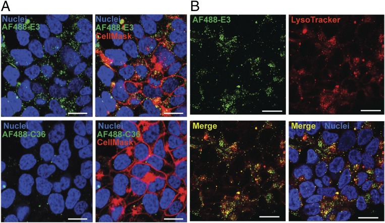
Fig. 3.
Confocal microscopy visualization of E3 internalization into 22Rv1 prostate cancer cells. Cells were treated for 1 h with 1 μM of either (A) AF488-E3 or AF488-C36 ± CellMask Deep Red Plasma Membrane Stain or (B) AF488-E3 + LysoTracker Deep Red stain to label acidic vesicles. After washing, Hoechst 33342 was added to all samples to stain the nuclei. Fluorescence was observed on a Leica SP5 inverted confocal microscope. (White scale bars: 20 μm.)

