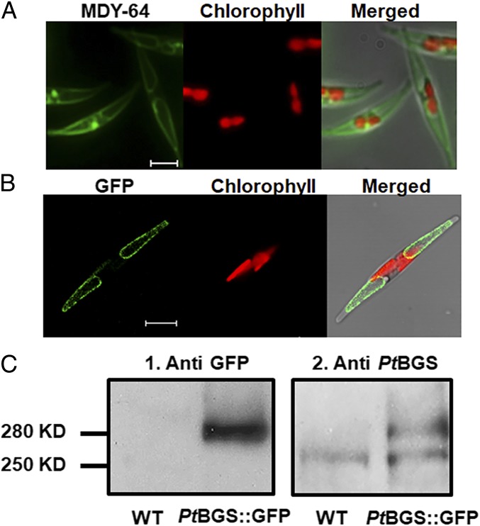
Fig. 1.
Localization of PtBGS in P. tricornutum. (A) MDY-64 stain of vacuoles in P. tricornutum. MDY-64 fluorescence (Left), chlorophyll fluorescence (Center), and a merged image (Right) are shown. (Scale bar: 5 μm.) (B) PtBGS::GFP fusion protein expressed in P. tricornutum. GFP fluorescence (Left), chlorophyll fluorescence (Center), and a merged image (Right) are shown. (Scale bar: 5 μm.) (C) Immunodetection of PtBGS::GFP in P. tricornutum. Western blots of proteins from WT and PtBGS::GFP lines (PtBGS::GFP) decorated with the antibodies against GFP (1) or PtBGS (2) are shown.

