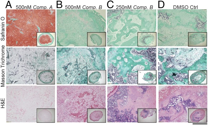
Fig. 5.
Development of hMSCs-derived cartilage tissues following 8 wk of ectopic implantation in vivo. Safranin O staining identifies GAG deposition; Masson Trichrome and H&E staining allow identify the presence of bone remodeling and bone marrow. (A) Compound A pretreatment was the only condition allowing the maintenance of a cartilaginous matrix still rich in GAG after 8 wk. (B–D) All of the other conditions exhibited a progression toward remodeling in bone. All images were taken at the same magnification (n = 3 donors). (Scale bar, 300 μm.)

