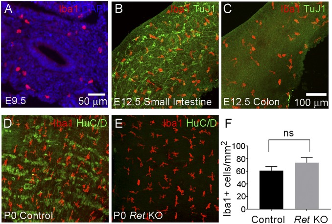
Fig. 1.
MM colonize the bowel with or without enteric neurons. (A) Sagittal section of WT E9.5 foregut stained with Iba1 antibody (red) and with DAPI (blue). This shows Iba1+ macrophages present in the bowel at E9.5. (B) WT E12.5 whole-mount small intestine stained with Iba1 (red) and TuJ1 (green) antibodies. (C) WT E12.5 whole-mount colon stained with Iba1 (red) and TuJ1 (green) antibodies. This shows Iba1+ macrophages in E12.5 WT colon that is not yet colonized by ENCDC. (D and E) Whole-mount P0 muscularis externa from control and Ret KO distal small intestine stained with Iba1 (red) and HuC/D (green) antibodies. Ret KO bowel is devoid of HuC/D+ enteric neurons, but has well patterned Iba1+ MM present in normal abundance. (F) Quantitative analysis of Iba1+ cell density in small bowel showed no statistically significant difference between WT and Ret KO small bowel (Student’s t test, P > 0.05, n = 8 mice per genotype). NS, not significant. Error bar = SEM. (Scale bar: 100 µm.) Scale bar in C applies to B–E.

