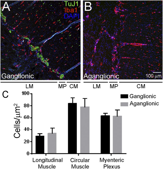
Fig. 2.
MM are present in normal abundance and distribution in human aganglionic colon from children with Hirschsprung disease. (A and B) Human colon was stained with antibodies to TuJ1 (green), Iba1 (red), and with the nuclear dye DAPI (blue). Iba1+ macrophages are present in ganglionic (A) and aganglionic (B) human muscularis externa in normal numbers and with normal distribution across muscle layers. (Scale bar: 100 µm.) (C) Quantitative analysis of MM abundance in layers of the muscularis propria. CM, circular muscle; LM, longitudinal muscle; MP, myenteric plexus. Student’s t test, P > 0.05 for all comparisons. n = 8 ganglionic and 8 aganglionic. Error bar = SEM.

