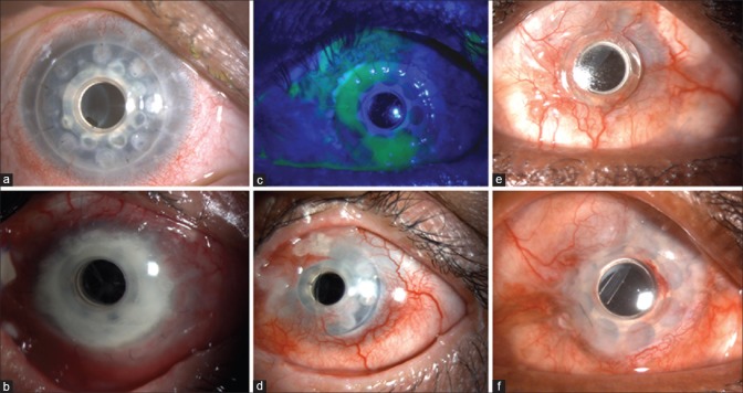
Figure 2.
(a) Retroprosthetic membrane in a silicone oil-filled eye after Boston Type 1 keratoprosthesis, not visually significant. (b) Carrier graft infiltration in an eye with vitreous exudates and endophthalmitis 2 years following Boston Type 1 keratoprosthesis. (c) Epithelial defect noted on fluorescein staining after BCL removal, not associated with thinning. (d) Sterile carrier graft melt with edge lift of the keratoprosthesis. (e) Perioptic annular melt with no leak. Note the air bubble in the gap beneath the flange of the optic. (f) Same eye as e following an annular lamellar graft

