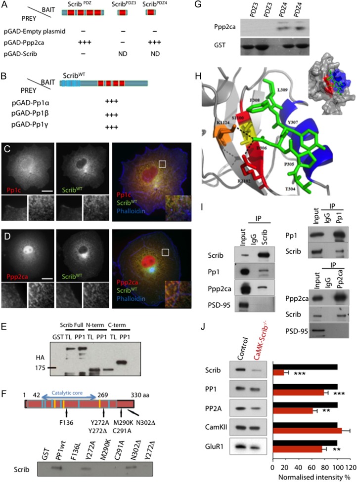
Figure 5.
Scrib scaffolds PP1 and PP2A phosphatases. (A) Directed yeast two-hybrid assays with Scrib and PP2A catalytic subunit (Ppp2ca) constructs. Schematic domain structures of ScribPDZ, ScribPDZ3, and ScribPDZ4 used as baits. Ppp2ca binds ScribPDZ and ScribPDZ4, but not ScribPDZ3. (B) Directed yeast two-hybrid assays with Scrib and Pp1 constructs. Pp1α,β,γ can bind Scrib. (C and D) ScribWT colocalizes with Pp1c (C) and Ppp2ca (D) catalytic subunits. COS-7 cells transiently co-transfected with hScrib-GFP (green) and HA-tagged Pp1c (red) (C) or hScrib-GFP (green) and Ppp2ca (red) (D). Phalloidin is shown in blue. Bottom white panels show higher magnification of individual or merged staining at the plasma membrane and cytoplasm level. Scale bars = 15 µm. (E) Three N-terminal HA-tagged constructs that contain either the full-length (Full) or N-terminal/LRR domain (N-term) or C-terminal/PDZ domains (C-term) truncated form of Scrib were expressed in COS cells. Lysates were pulled down with GST alone (GST) or GST-PP1 (PP1) and submitted to SDS-PAGE and western blot analysis with an anti-HA antibody. Input lanes (TL) represent 10% of total extract used for each pull down; (F) Schematic representation of PP1: the catalytic core domain span from amino-acid residue 42–269 and the binding domain from 270 to 330. Blue and Yellow stripes represent GDxHG, GDxVDRG or GNHE sequences. The different point mutations (F136L, Y272 A, M290K, C291A) and truncations of the tyrosine in position 272 [Y272Δ] or asparagine in position 302 [N302Δ] are mentioned with their corresponding positions. Resulting complexes were analyzed by anti-Scrib antibody western blot. (G) GST pull downs indicate that Ppp2ca can interact with PDZ4 in a dose-dependent manner but not with PDZ3. (H) Model of the interaction of Scrib-PDZ4 K1124 (orange) and R1102 (red) residues with the T304 to L309 residues from Ppp2ca C-terminus (green) and overall view of the model of the interaction (top right) between the alpha helix B (blue) and beta strand B (red) of Scrib-PDZ4 (gray) and Ppp2ca C-terminus (green). (I) Endogenous coimmunoprecipitation of Scrib, Pp1 and Ppp2ca from the hippocampus. Supernatants were immunoprecipitated with Scrib antibodies. The precipitates show positive immunoblotting for Pp1 and Ppp2ca subunits. (J) Immunoblot analysis of PSD fractions from Control and CaMK-Scrib−/− mice, and quantification of Scrib, CaMKII, PP1 and PP2 proteins. The blots were normalized to WT (100%; black histograms). Data were compared using Mann–Whitney, P < 0.01. Data are represented as mean ± SEM.
The following figure supplement is available for Figure 5: Supplementary Fig. S4. CaMK-Scrib1−/− mice exhibit normal level of PP1, PP2A, GluR1, and PSD95 in cell lysates.

