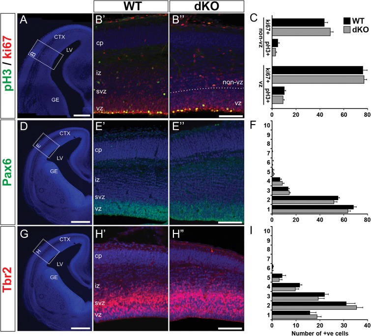
Figure 2.
Absence of AU040320 and KIAA0319 does not alter cortical neurogenesis. (A, D, G) Representative DAPI images outlining regions of E15 cortices selected for analysis. (B) Immunolabeling of cycling cells with ki67 (red) and cells in M-phase with pH3 (green) to examine cell division profile in WT (B’) and dKO mice (B”). (C) Quantification of number of ph3+ and ki67+ cells in the VZ region and in the rest of the cortical wall (non-VZ); no differences were observed between the 2 genotypes (n = 3, P > 0.05). (E, H) Pools of neuronal progenitors were examined by labeling radial glial (Pax6+, green; E) and intermediate progenitors (Tbr2+, red; H). (F, I) Quantification of number of cells in each of the 10 equally sized bins dividing the cortex (in E, H) revealed no differences between dKO and WT sections for any of the conditions (n = 3, P > 0.05). All image panels show nuclear staining with DAPI. All data shown as mean ± SEM. CTX, cortex; LV, lateral ventricle; GE, ganglionic eminence; cp, cortical plate; iz, intermediate zone; svz, subventricular zone; vz, ventricular zone. Scale bars: 400 μm (A, D, G); 100 μm (B, E, H).

