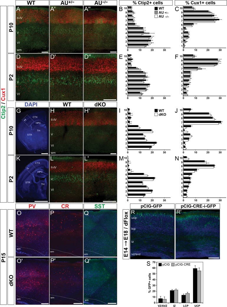
Figure 3.
Normal cortical lamination in AU040320 and AU040320;Kiaa0319 KO brains. (A–F) Immunohistochemistry labeling lower layer pyramidal cells (V–VI, Ctip2; green) and upper layer ones (II–IV, Cux1+; red) in somatosensory cortex (S1) of AU040320 mutants at P10 (A) and P2 (D). Quantification graphs for percentage of Ctip2+ and Cux1+ cells per bin at each age (B, C for P10; E, F for P2) show no differences across each condition (n = 3, P > 0.05). (G–N) Similar analyses were conducted for dKO brains. DAPI-stained images (G, K) show the cortical region (S1; dotted lines, insets) selected for quantification, for both AU040320 and double KOs. Graphs with quantification of cell distribution per bin (I, J for P10; M, N for P2) indicate that absence of AU040320 and KIAA0319 leaves cortical lamination unaffected for all conditions (n = 3, P > 0.05). (O–Q) Three subpopulations of interneurons were examined for their overall distribution in the S1 cortex of dKO brains at P15: parvalbumin+ (PV, red; O) cells, calretinin+ (CR, red, P) and somatostatin (SST, green, Q). No differences were detected in any of the conditions (n = 3). (R, S) In utero electroporation of plasmids expressing GFP only (pCIG-GFP, R) and Cre recombinase with GFP (pCIG-CRE-i-GFP, R’) into the brains of E14 double Kiaa0319;AU040320 floxed mice (dFlox) which were harvested at E18. Quantification of distribution (%) of cells in each subdivision of the cortical wall reveals no differences following the acute knockout of Kiaa0319 and AU040320 combined (S; n = 3, P > 0.05). Image panels O–R show nuclear staining with DAPI. All data shown as means ± SEM. AU, AU040320; wm, white matter; ucp, upper cortical plate; lcp, lower cortical plate; iz, intermediate zone; svz, subventricular zone; vz, ventricular zone; CTX, cortex; HPC, hippocampus; STR, striatum; TH, thalamus. Scale bars: 75 μm (A, D, H, L); 1000 μm (G, K); 150 μm (O–R).

