Figure 3.
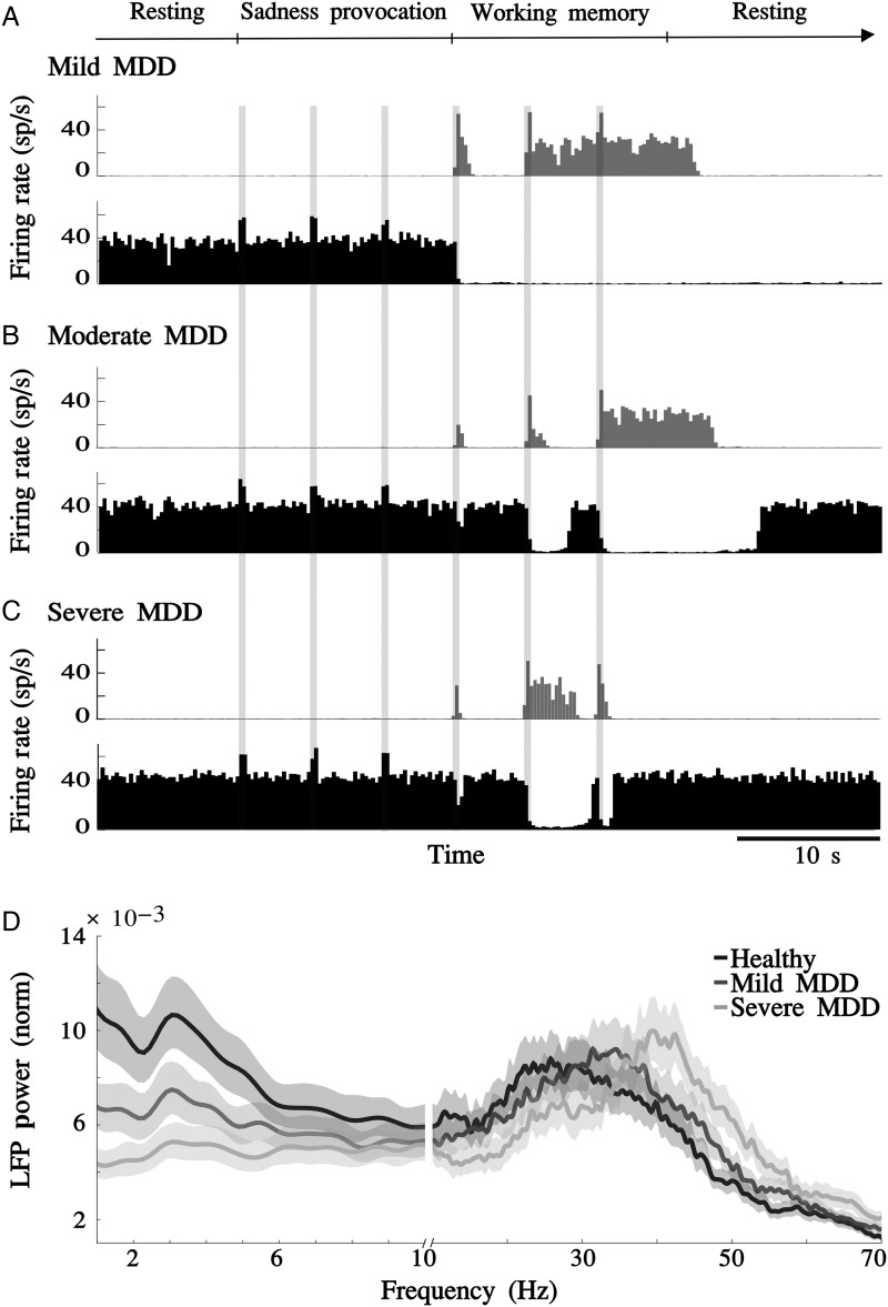
Glutamate decay slow-down reproduces the progressive nature of MDD. (A) Mild MDD network (2.5% slow-down in glutamate decay in the vACC). Activity histograms for a single simulation show aberrant activity in the vACC during the resting epoch. Although dlPFC only responded partially to the first inputs, it was still able to turn off activity in the vACC during the WM epoch. (B) Moderate MDD network (5% slow-down in glutamate decay in the vACC). vACC showed aberrant activity in resting epochs and dlPFC showed diminished responsivity to cognitive inputs, now being unable to turn off vACC. (C) Severe MDD network (7.5% slow-down in glutamate decay in the vACC). Aberrant vACC activity was not modulated by any kind of inputs. (D) LFP normalized power spectrum in the vACC. Synchronization in the theta frequency range was progressively reduced, whereas beta/gamma rhythms were enhanced as glutamate decay was gradually slowed down (healthy, 2.5% and 7.5% slow-down in glutamate decay). Jackknife error bars around the mean mark the 95% CI.

