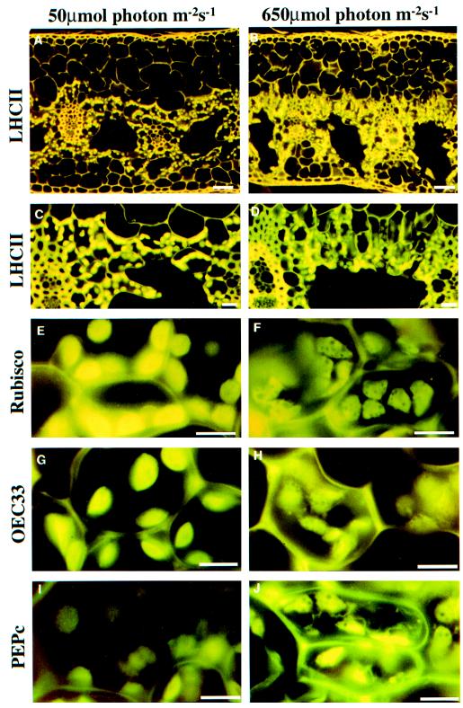
Figure 1.
Immunolocalization of PEG-embedded G. monostachia leaf sections (10 μm thick). A, LL at low magnification; B, HL at low magnification; C, LL at high magnification; and D, HL at high magnification. Leaf sections were incubated with primary antisera to LHCII (A–D), Rubisco (E and F), OEC33 (G and H), and PEPc (I and J) followed by secondary goat anti-rabbit antisera conjugated to fluorescein isothiocyanate for LL plants (A, C, E, G, and I) and HL plants (B, D, F, H, and J). The scale bars represent 50 μm (A and B), 20 μm (C and D), and 10 μm (E–J).

