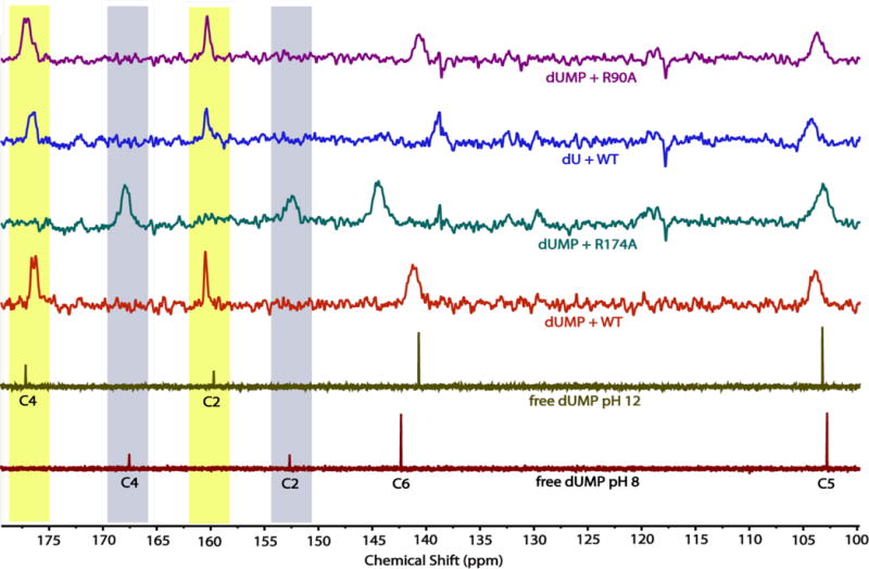
Figure 4.

The 13C-NMR spectra of uracil carbons of dUMP/dU free in solution or bound to WT and variant FDTSs at 45 °C. All of the ligand-FDTS complexes were at pH 8. The C4 and C2 signals for dUMP shifted upfield ~8 ppm when bound to WT FDTS, having nearly identical chemical shifts as free dUMP at pH 12. Mutagenesis of R174 – which is 2.8 Å from N3 of dUMP in the dUMP-WT FDTS structure – to alanine eliminated the changes in chemical shifts of the dUMP-FDTS complex. In contrast, mutagenesis of R90 to alanine or removal of the phosphate of dUMP had little effect on the 13C-NMR spectrum of the uracil carbons in the complex. Highlighted in maize and blue, respectively, are the chemical shifts of the carbonyl carbons of N3-ionized and N3-unionized uracil.
