Fig. 2.

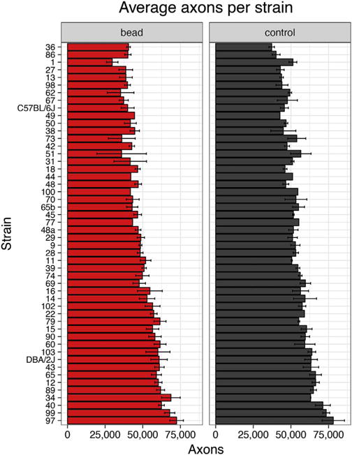
Axon counts for the normal eye (‘control’, grey) and for the eye 21 days after elevation of IOP (‘bead’, red). Error bars show standard error.

Axon counts for the normal eye (‘control’, grey) and for the eye 21 days after elevation of IOP (‘bead’, red). Error bars show standard error.