. 2018 Mar 19;9(15):3782–3787. doi: 10.1039/c8sc00429c

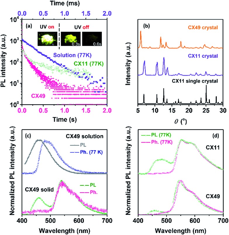
Fig. 2. (a) Time-resolved emission decay curves of CX under different conditions: CX49 crystal (545 nm), CX11 crystal (545 nm, 77 K) and in toluene solution (485 nm, 10–5 M, 77 K, top coordinate). Inset photos are the CX49 crystal sample before (on) and after (off) the UV-light exposure removal. (b) Comparison of the powder X-ray diffraction spectra of CX49 and CX11 crystals, and the simulated spectrum from the CX11 single crystal. (c) Steady-state photoluminescence (PL) spectra and phosphorescence spectra (Ph, delayed 8 ms) of CX in toluene solution (10–5 M) and the crystalline (CX49) state. The spectra were recorded at room-temperature unless otherwise stated. (d) Steady-state PL spectra and Ph spectra (delayed 8 ms) of CX11 and CX49 crystals at 77 K.

Fig. 2