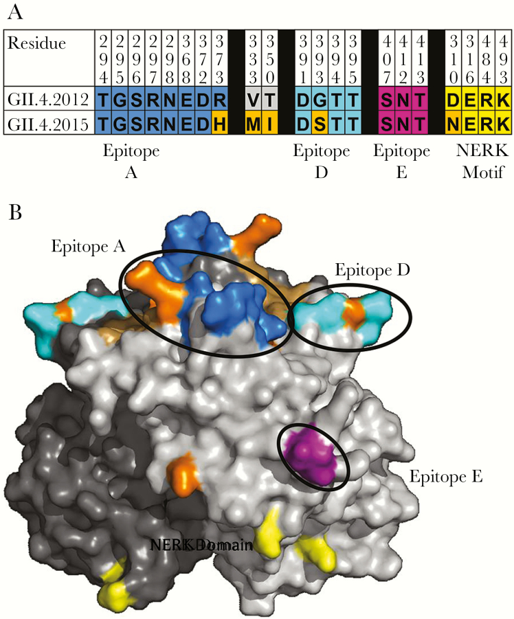
Figure 2.
Capsid residues defining antigenic and ligand-binding sites differ between GII.4 2012 and GII.4 2015. A, GII.4 2012 and GII.4 2015 differ at 5 residues in the P2 subdomain of the major capsid protein (orange). B, Characterized blockade antibody epitopes and residue changes between GII.4 2012 (JX459908) and GII.4 2015 (KX907727) were highlighted onto the P domain dimer of GII.4 2012 [34] (residue 333 is not visible in this view). Residues within epitopes A (blue) and D (cyan) and the NERK motif (yellow) of the major capsid protein differ by single amino acid changes between GII.4 2012 and GII.4 2015 (orange). The carbohydrate-binding domain remains conserved (brown).

