Figure 1.
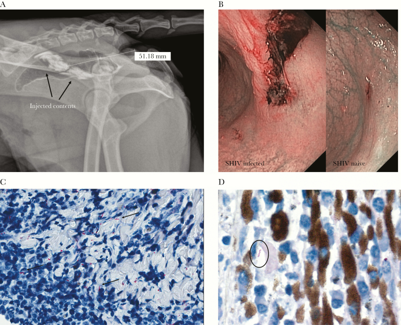
A nonhuman primate model for rectally acquired syphilis. A, Radiograph showing the location and dispersion of submucosally injected contents in the rectum of rhesus macaques (2 injections in a 0.2-mL volume; animal 1 in Table 1). Radiography was done separately, not by mixing the contrast medium with Treponema pallidum. The injections were administered approximately 5 cm beyond the anorectal opening. B, Narrow-band imaging showing representative pictures of ulcerated rectal lesions in the rectum of simian/human immunodeficiency virus (SHIV)–infected (day 7; animal 2) and SHIV-naive (day 6; animal 4) macaques. C, Immunohistochemical staining using a commercial rabbit polyclonal antibody raised against Borrelia burgdorferi showing T. pallidum dissemination within lymphoplasmacytic conjunctival infiltrates of animal 1 (400× original magnification). D, Immunohistochemical staining using T. pallidum–specific rabbit polyclonal antibody showing T. pallidum dissemination within lymphoplasmacytic infiltrates in the ciliary body of the eye of animal 1 (630× original magnification). In contrast to biopsy specimens from back lesions, none of the biopsy specimens from rectal lesions showed the presence of T. pallidum by polymerase chain reaction analysis or immunohistochemical staining. We believe this reflects the challenge of sampling specific sites in the macaque rectum without proper visualization.

