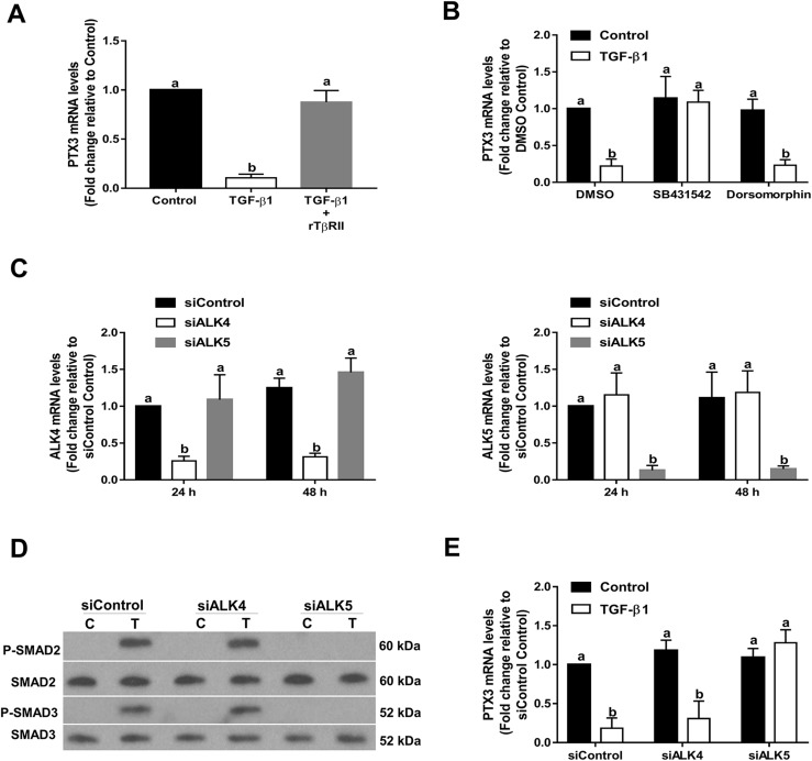
Figure 3.
TGF-β1 downregulates the expression of PTX3 via a TβRII/ALK5-mediated signaling pathway in SVOG cells. (A) A total of 5 µg/mL rTβRII was preincubated with 10 ng/mL recombinant TGF-β1 at room temperature for 1 hour and was then added to SVOG cells for 24 hours. PTX3 mRNA levels were examined using RT-qPCR. (B) Cells were treated with 10 ng/mL TGF-β1 for 6 hours in the presence of vehicle control [dimethyl sulfoxide (DMSO)], 10 μM SB431542 or 10 μM dorsomorphin dihydrochloride. PTX3 mRNA levels were examined using RT-qPCR. (C) Cells were transfected with 25 nM siRNA [small interfering (si)Control, siALK4, or siALK5] for 24 or 48 hours, and the mRNA levels of ALK4 or ALK5 were examined using RT-qPCR. (D) Cells were transfected with siRNA (siControl, siALK4, or siALK5) for 48 hours and then treated with 10 ng/mL TGF-β1 for 45 minutes. Phosphorylation levels of SMAD2 and SMAD3 were examined using western blot analysis. (E) Cells were transfected with siRNA (siControl, siALK4, or siALK5) for 48 hours and then treated for an additional 6 hours with vehicle control or 10 ng/mL TGF-β1. PTX3 mRNA levels were examined using RT-qPCR. The results are expressed as the mean ± standard error of the mean of at least three independent experiments; lowercase letters indicate statistically significant differences (P < 0.05). C, control; P, phosphorylated; T, TGF-β1.

