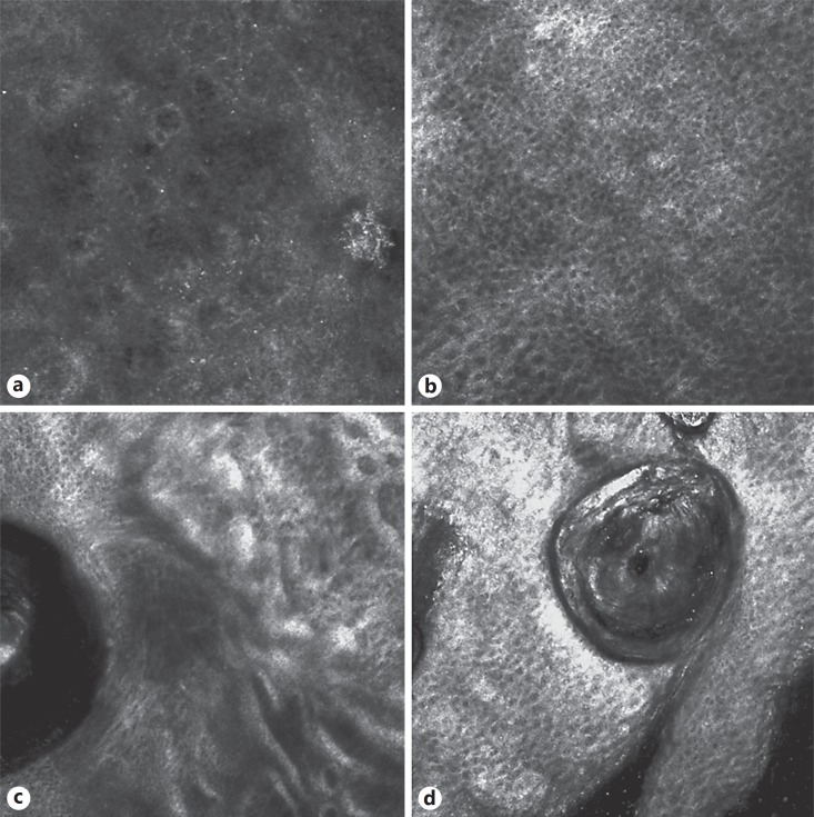
Fig. 2.
Confocal microscopy presentation: an infiltrate of small weakly refractile round to oval cells in the epidermis (a) with disorganized honeycomb pattern (b); refractile filamentous thick structures around the follicular structure (c); refractile round to oval cells of the remaining infundibular follicular structure (d).

