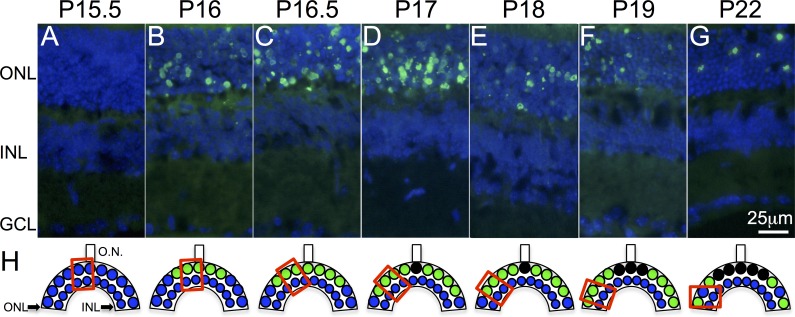
Figure 1.
Photoreceptor cell death in rd10 mouse retina. (A, B) PR cell death (green nuclei) starts abruptly at P16 in the central retina, with no apoptotic nuclei detected neither at P13.5 (data not shown) nor at P15.5. (C–G) Cell death progresses toward the periphery. (H) Schematic representation of the PR cell death process. Preapoptotic nuclei are represented by blue circles and apoptotic nuclei are represented by green circles. Red squares represent the retinal areas where images were captured. Cell nuclei (blue) are labeled with 4′,6-diamino-2-fenilindol (DAPI), GCL, ganglion cell layer; INL, inner nuclear layer; ONL, outer nuclear layer; ON, optic nerve.

