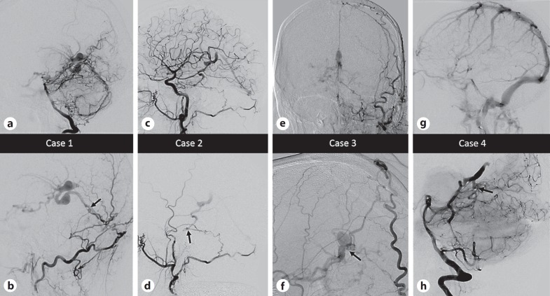
Fig. 2.
Select angiographic images for the patients described. In the lower row, the black arrows point to the location of the fistulous connection. In all cases multiple arterial feeders from both dural and parenchymal vessels were present, and select images are shown to best illustrate anatomical considerations. a, b Case 1. a Lateral angiogram, right vertebral artery injection, mid-arterial phase demonstrating enlargement of the posterior meningeal artery and artery of the falx cerebelli feeding a dural fistula with drainage into variceal superior hemispheric veins and the vein of Galen. b Lateral magnified angiogram, left occipital artery injection, mid-arterial phase demonstrating transosseous supply and venous fistula anatomy in greater detail. c, d Case 2. c Lateral angiogram, right common carotid injection, mid-arterial phase showing right meningohypophyseal trunk and right occipital artery supply to a point of fistulation along the right petrous apex. Variceal venous structures are then seen to fill in the arterial phase, including faint opacification of the straight sinus. d Lateral angiogram, right external carotid injection, mid-arterial phase showing transosseous dural supply from the occipital artery isolating the point of fistulation. e, f Case 3. e Anteroposterior angiogram, left external carotid injection, late arterial phase showing right transosseous superficial temporal artery, middle meningeal artery, and occipital artery feeders sending dural falcine branches deep to the point of fistulation adjacent to the vein of Galen, which is variceal and seen to fill in the arterial phase. f Lateral magnified view, right external carotid injection, late arterial phase showing similar anatomy on the contralateral side with large dural falcine branches and point of fistulation on the lateral wall of the vein of Galen. g, h Case 4. g Lateral angiogram, left internal carotid artery injection, venous phase showing absence of deep venous structures. The structures did not opacify in the venous phase on any arterial injections, due to a combination of arterialized flow and thrombosis. h Lateral magnified angiogram, right vertebral injection demonstrating a midline fistula fed by bilateral superior cerebellar arteries with drainage into a dilated basal vein of Rosenthal and retrograde into lateral pontomesencephalic veins. The vein of Galen was thrombosed and did not opacify.

