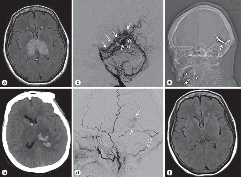
Fig. 3.
a Initial axial FLAIR MRI showing bilateral thalamic hyperintensity. b Axial CT showing hemorrhagic change within the congested tissue of the left thalamus with intraventricular extension. c Digital subtraction angiography right vertebral injection, lateral view showing enlargement of the posterior meningeal artery and artery of the falx cerebelli feeding a dural fistula (open arrow) with drainage into variceal deep venous structures (white arrows) that fill in the arterial phase. d Digital subtraction angiography, left external carotid injection, lateral view showing left middle meningeal artery feeder to the same point of fistulation (open arrow), with similar venous egress (white arrows). e Scout image from post-procedural CT showing Onyx cast following deposition in the middle meningeal supply successfully occluding the point of fistulation (white arrow). f FLAIR MRI 3 months post-procedure showing slowly improving deep tissue changes.

