FIGURE 4.
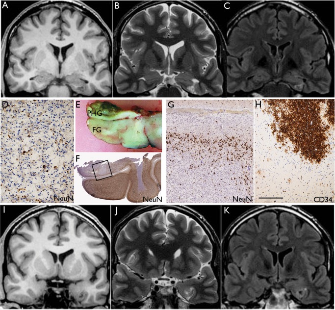
Diffuse GNT with mixed features of DNT. Case 21: A lesion centered in the left parahippocampal gyrus. (A) Coronal T1, (B) T2, and (C) FLAIR MR images showing the lesion involving the left parahippocampal gyrus with a triangular wedge-shaped appearance and a multiple cystic appearance. The wedge-shaped lesion points toward the temporal horn of the left lateral ventricle and the appearance of the tumor in this region resembles the complex subtype of DNT. (D) Tissue from the main lesion showed glioneuronal element with floating neurons characteristic of DNT on NeuN stain. (E) In the main temporal lobe specimen, which included the fusiform gyrus (FG) and part of the parahippocampal gyrus (PHG), there was diffuse pattern of tumor infiltration which was the dominant overall growth pattern as shown on NeuN stain in (F). The region shown in the square is shown at higher magnification in (G) and rotated 180°. (H) The infiltrating cells showed patchy CD34 labeling. (I) Coronal T1, (J) T2, and (K) FLAIR corresponding to the level shown in (E) confirmed an ill-defined white matter signal abnormality with cortical atrophy; this part of the lesion resembled diffuse GNT. Bars: D = 100 μm; G, H = ∼175 μm, based on original magnifications.

