. 2018 Apr 3;9(15):3820–3827. doi: 10.1039/c8sc00074c

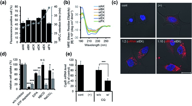
Fig. 2. (a) Cell penetrating abilities of 10 nM of stapled peptides (mean ± s.d., n = 3) and their HPLC retention times in gray. (b) CD spectra of 20 μM of stapled peptides in 10 mM sodium phosphate (pH 7.4). (c) Confocal laser scanning microscopy (CLSM) images of HeLa cells transfected with 50 nM of DY-547 labeled siRNA (siGLO™) (red) for 24 h. Cell nuclei were stained with Hoechst 33342 (blue). DharmaFECT™ 1 was used as a positive (+) transfecting agent (scale bar, 20 μm). (d) Relative cell penetration of fluorescence labeled stEK (black) and stEK-siGLO™ complex (gray) in the presence and absence of endocytosis inhibitors (mean ± s.d., n = 3, N.S. not significant, *P < 0.5, **P < 0.01, ***P < 0.001 compared with the control w/o inhibitor). ATP depletion, 5-(N-ethyl-N-isopropyl)amiloride (EIPA) and sodium chlorate (NaClO3) were used to block the ATP-dependent pathway, macropinocytosis and proteoglycan-dependent endocytosis, respectively. Methyl-β-cyclodextrin (MβCD) was used to inhibit cholesterol associated membrane processes, including caveloin, lipid raft, and macropinocytosis. (e) In vitro gene silencing efficiency of siRNA–stEK complexes in HeLa cells. Cells were incubated with 50 nM of siRNA and 500 nM of stEK in the presence and absence of 120 μM of chloroquine (CQ). Relative mRNA expression levels were determined by RT-qPCR (mean ± s.d., n = 3, ***P < 0.001 compared with the control w/o CQ).

Fig. 2