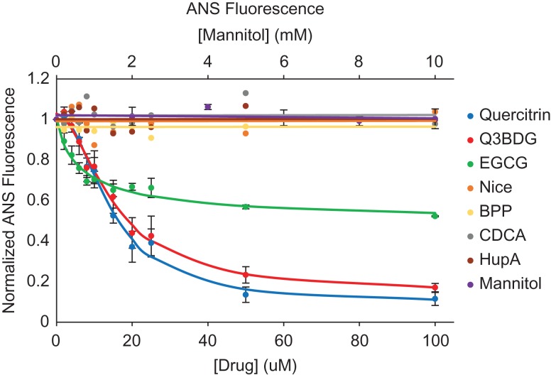
Fig. 3.
A4V SOD1 oxidation-induced misfolding and its inhibition with quercitrin, Q3BDG, and EGCG. A4V SOD1 was misfolded using H2O2 at room temperature for 24 h in the presence and absence of increasing concentrations of the various drugs. Following this time period, ANS was added as a fluorescent dye that binds to exposed hydrophobic pockets where higher fluorescence indicates misfolding. The resultant fluorescence was normalized to the highest (no drug) and lowest (no H2O2) signals using Equation (3). The averages of three replicates are graphed here while error bars representing the standard error of the mean are shown for quercitrin, Q3BDG, EGCG, and mannitol. The EC50 values can be found in Table I.

