Figure 3.
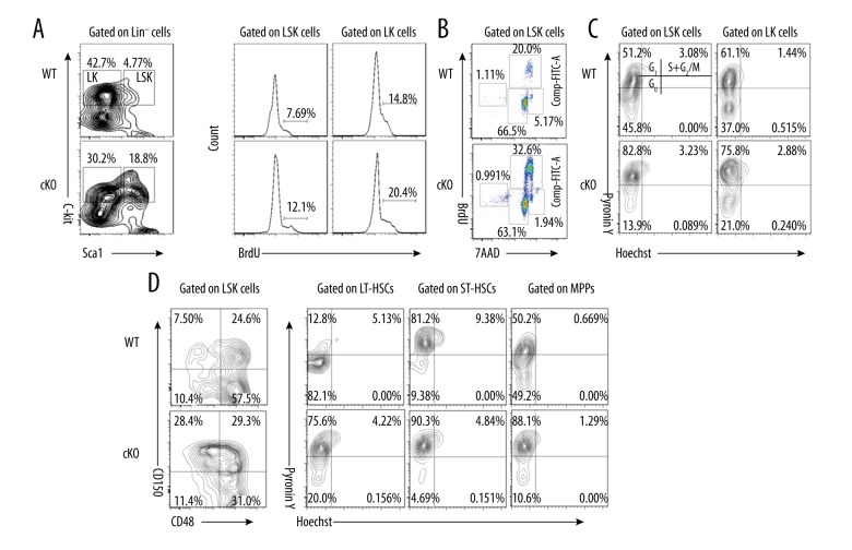
Loss of MYSM1 drives HSCs from quiescence to rapid proliferation. (A) WT and cKO mice received 2 mg BrdU intraperitoneally daily for 5 days. Incorporation of BrdU was analyzed by FACS in BM LSK and LK cells (n=4 per group). (B) Mice received 2 mg BrdU i.p. injection 1 hour before sacrifice. BM cells were isolated and stained for cell cycle analysis (n=4 per group). (C, D) WT and cKO BM cells were stained for HSC surface antigens followed by Hoechst 33258/Pyronin Y staining. Representative FACS plots of cells depicting G0 (bottom left quadrant), G1 (top left quadrant), and S/G2/M (top right quadrant) in (C) LSK and LK cells and (D) LSK subsets (n=4 per group).

