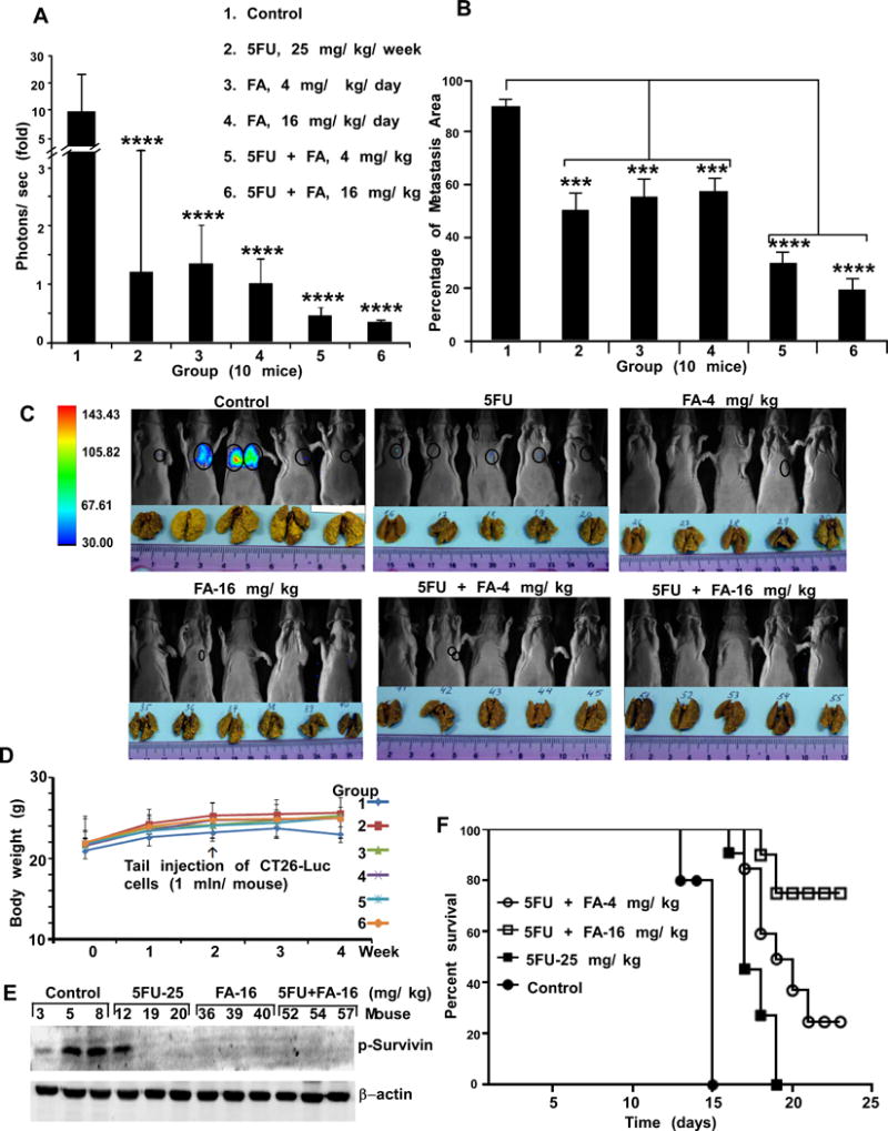
Figure 5.

FA alone or combined with 5-FU suppresses colon lung metastasis in mice. A, CT26-Luc cells were injected into the tail vein (n = 10 mice/group). Lung tumors were observed by in vivo optical imaging with D-luciferin, which was injected intraperitoneally. Before inoculating CT26-Luc cells, groups 3-6 were intraperitoneally injected with FA for 10 days. The imaging was performed using the Xtreme Image system and bioluminescence was quantified using Bruker MI. The asterisks (****) indicate significantly less metastases (p < 0.001) in CT26-Luc cells in treated mice. B, lungs were fixed in Bouin’s solution to visualize metastatic nodules (yellow color) and data were analyzed using GraphPad Prism 5. The percentage of lung metastasis observed in treated groups was significantly decreased (***p < 0.01, ****p < 0.001) compared with untreated controls. The treated groups of mice have the same nomenclature as panel A. C, representative mice (5 mice/group) with bioluminescence signal (circled) and metastatic lungs (yellow color) are shown. D, FA or its combination with 5-FU does not affect mouse body weight. The treated groups of mice have the same nomenclature as panel A. E, the expression levels of phosphorylated survivin were analyzed in lung tissues (3 mice/group) by Western blotting. F, overall percent survival of animals (9 mice/group) with experimental lung metastasis of CT26-Luc colon cancer cells after treatment with 5-FU, FA or the combinations 5-FU and FA.
