Figure 1.
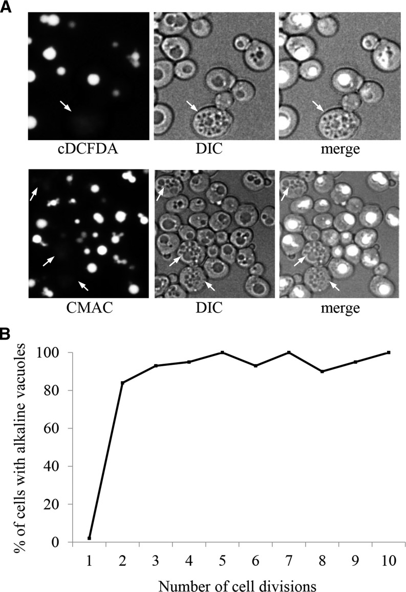
Vacuolar morphology and acidity change as yeast cells age. (A) WT cultures grown overnight were stained with the pH sensitive vacuolar fluorescent probes CMAC-Arg or carboxy-DCFDA (cDCFDA). Both stains fluoresce when subjected to an acidic environment. Cells were grown and stained at physiological pH. Arrows denote representative older cells with fragmented vacuoles that lack staining, indicative of alkalized vacuoles. See Figure S1, A and B showing that yeast cells with greater than 4 buds scars typically display fragmented vacuoles. (B) Populations were enriched for older cells using the Mother Enrichment Program (MEP; Lindstrom and Gottschling 2009). Cells were binned according to their indicated number of bud scars and scored as having alkaline vacuoles if a twofold or greater reduction in fluorescence intensity was observed compared to young cells with one bud scar. Note that the plot reaches a steady state in cells with greater than 3 divisions. The bins contain anywhere between 20 (for cells with high number of bud scars) and 40 (for cells with few number of scars) cells. See Figure S1C for cDCFDA fluorescence emission spectra of young and old yeast.

