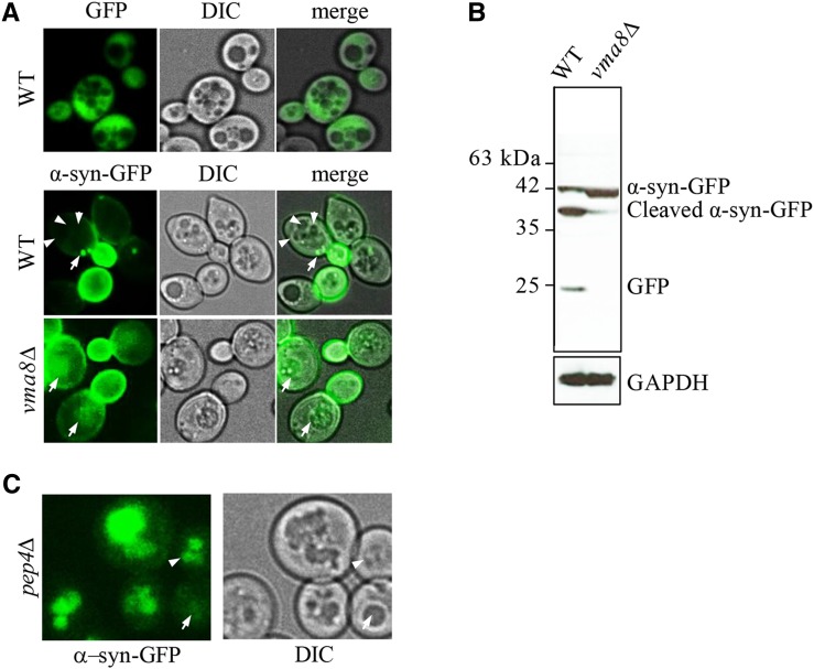
Figure 3.
Cells with impaired vacuolar function accumulate uncleaved α-syn-GFP aggregates within their vacuoles. (A) Live cell confocal imaging of genomically integrated α-syn-GFP expressed from a GAL1-inducible promoter in WT and vma8Δ mutants grown in YP + 0.1% galactose. Expression of a GFP reporter using the GAL1 promoter results in cytoplasm fluorescence (top panel). Arrows denote cytoplasmic GFP foci and arrowheads point to vacuolar lumen in WT cells. See Figure S3A for images of α-syn-GFP aggregation as galactose concentrations increase. (B) GFP westerns in whole cell lysates prepared from cells in (A). GAPDH served as a loading control. (C) Live cell imaging of dividing pep4∆ cells expressing genomically integrated α-syn-GFP induced from a GAL1 promoter. Cells were grown in 0.1% galactose. The arrow depicts a very young cell with subtle vacuolar staining; the arrowhead shows a young cell experiencing vacuolar fragmentation with marked vacuolar staining.

