Figure 3.
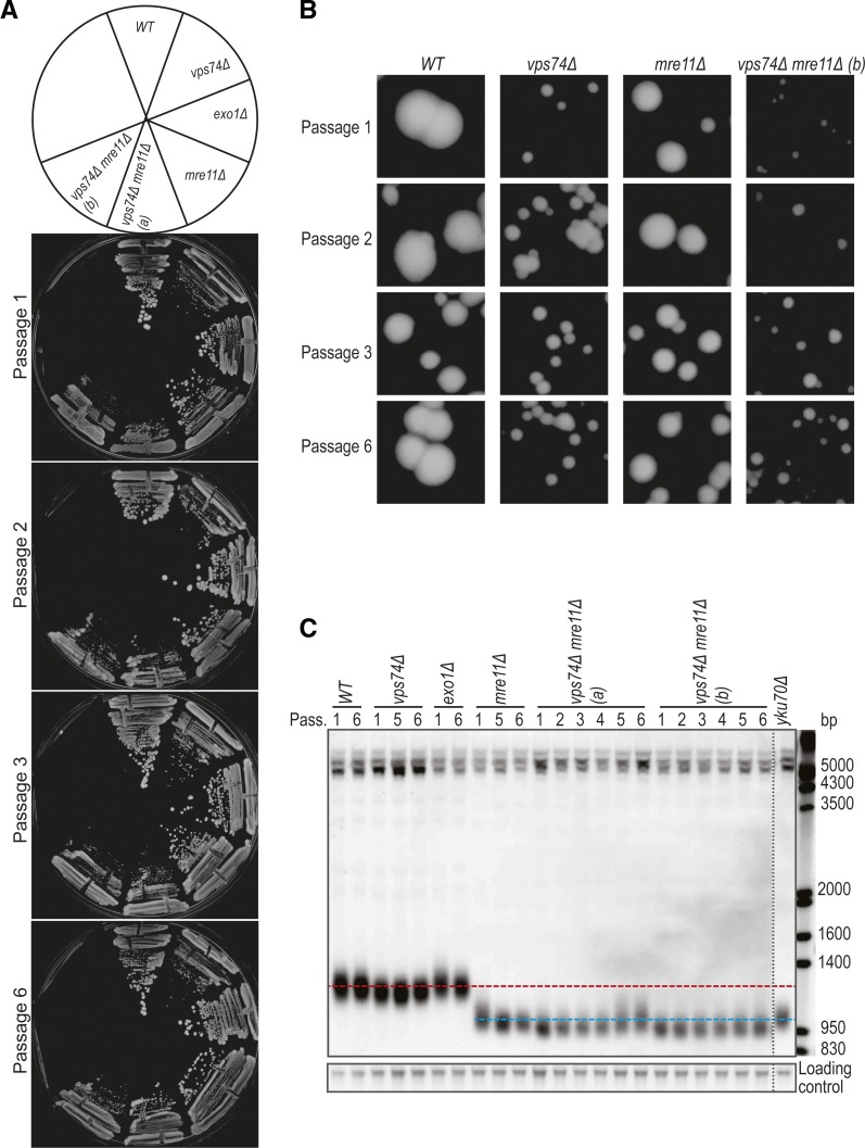
Loss of VPS74 leads to telomere shortening in WT and mre11Δ cells. (A) Passage tests performed at 30°C. Cells were allowed to grow for 2 days before pictures were taken and cells passaged. (B) Zoom in of the colonies in A. (C) Cells from the plates in A were inoculated in liquid YEPD, grown until saturation and DNA was extracted. The DNA was analyzed by Southern blot with a telomere probe (Y’+TG). Horizontal red line represents the WT telomere length and the blue line is roughly the telomere length of mre11Δ cells. Vertical dashed line indicates where the gel picture was cut for presentation purposes.

