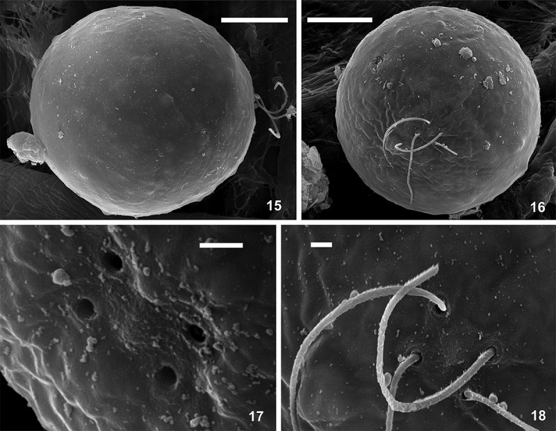
Figs 15–18.

SEM micrographs of Chlainomonas sp. swarmers from the snow of the Ľadové Lake. Figs 15, 16. Side and apical view showing the smooth surface of quadriflagellate cells. Fig. 17. Detail view of four spherical flagellar grooves in slightly rectangular position. Fig. 18. Detail view of two pairs of flagella. Scale = 10 µm (Figs 15, 16) and 1 µm (Figs 17, 18).
