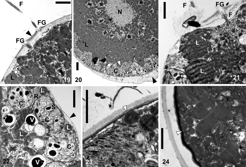
Figs 19–24.

TEM micrographs of Chlainomonas sp. swarmers (Figs 19–22) and spores (Figs 23, 24) from the snow of the Ľadové Lake. Figs 19, 20. A section showing flagellate (F) and two flagella grooves (FG) of a swarmer. Cell wall thickened at the anterior and the posterior of the cell (black arrows). Note small chloroplasts (C) located parietally and a putative process of plastid division (white arrow). Centrally located nucleus (N), likely surrounded by many lipid bodies (L). Figs 21, 22. A swarmer with a single, equally thin cell wall (black arrow) with papilla. Section showing one flagella (F) and the flagella groove (FG). Note lipid bodies (L), starch grains (S) in chloroplasts (C), electron dense vacuoles (V) containing a crystalline content and a ribosome rich region (R) close to the cell wall. Figs 23, 24. Spherical spore with the trilaminar sheath (secondary cell wall, white arrow). Later outer layers of extracellular matrix are developed. Scale = 2 µm.
