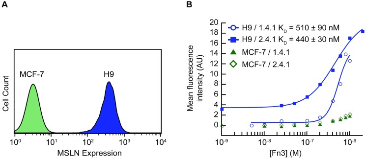
Fig 5. Engineered Fn3 protein variants bound cancer cells expressing MSLN.
A431/H9 cells, epidermoid carcinoma cells transfected to express high levels of MSLN, and MCF-7 cells, breast cancer cells lacking surface MSLN, were used in all binding assays. (A) Analysis by flow cytometry confirms MSLN presence on the surface of A431/H9 cells as detected by an anti-MLSN antibody. The MCF-7 cell line does not express MSLN. (B) Fn3 variants 1.4.1 and 2.4.1 were isolated and binding to MSLN was measured using equilibrium binding assays. A431/H9 and MCF-7 cells were incubated with a range of concentrations of soluble fluorescently labeled 1.4.1 or 2.4.1. The assays were performed in experimental triplicate. Data from each replicate were fit to a sigmoidal curve, and a KD value was calculated for each replicate. The KD is reported as the mean +/- standard deviation. A representative binding curve of each clone for both cell lines is shown.

