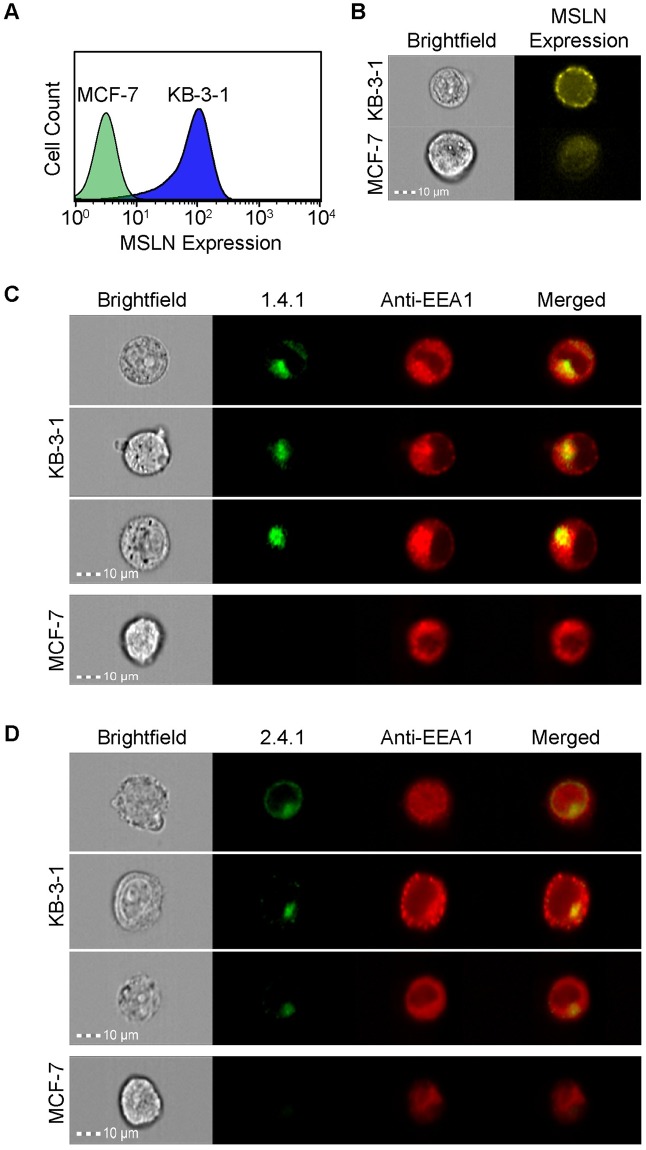
Fig 6. Engineered Fn3 protein variants 1.4.1 and 2.4.1 localized to early endosomes upon binding MSLN.
Analysis by (A) flow cytometry and (B) imaging flow cytometry confirms MSLN presence on the surface of KB-3-1 cells compared to the MSLN-negative MCF-7 cells, as detected by an anti-MSLN antibody. (C, D) KB-3-1 cells (top) internalize AF488-labeled 1.4.1 (C) and AF488-labeled 2.4.1 (D), while MCF-7 cells show no specific binding or internalization (C bottom, D bottom). Endosomes are detected by an AF647-conjuated antibody recognizing the EAA1 early endosomal marker. Yellow in the merged images indicate co-localization between AF488-1.4.1 or AF488-2.4.1 anti-MSLN engineered proteins (green) and EEA1 (red). Original magnification 40X. Co-localization is quantified by the Bright Detail Similarity (BDS) metric, with values near 1 indicating co-localization. KB-3-1 BDS = 1.31 and 0.919 for AF488-1.4.1 and AF488-2.4.1, respectively. BDS values are not quantifiable for the negative control cell line, due to insufficient fraction of negative control cell population staining for binding or internalization of engineered protein variants.

