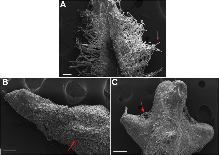
Fig 5. Scaning electron micrograph of Red Araza (Psidium cattleianum) roots.
(A) Control root, arrow shows root hairs. (B) and (C) Ectomycorrhizae formed by symbiotic relationship with the D17 isolate (Pisolithus microcarpus), arrow shows thick fungal mantle. Bars on A and B represent a 100 μm scale and, on C, a 200 μm scale.

