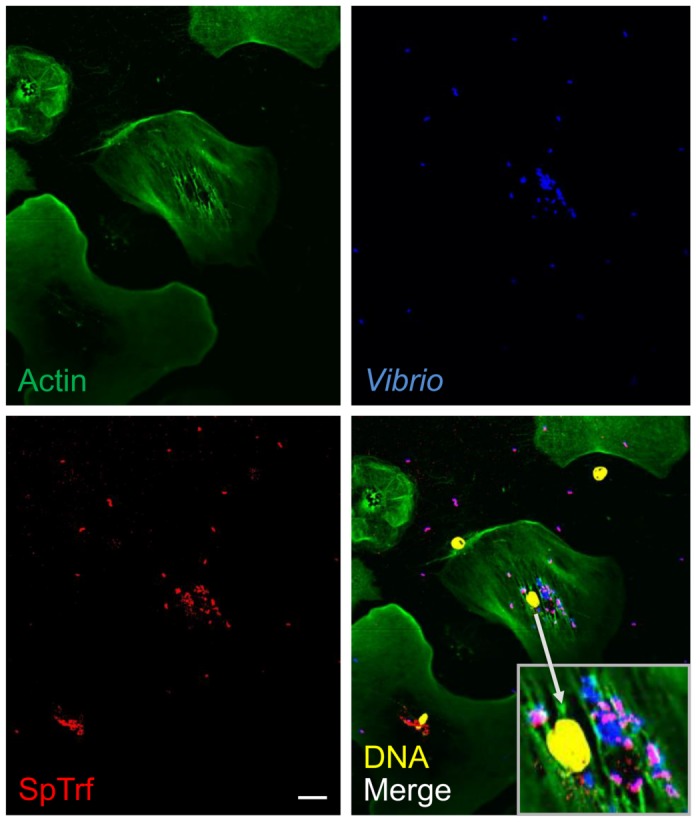
Fig 7. SpTrf proteins are secreted from coelomocytes in culture and opsonize Vibrio diazotrophicus.

Non-opsonized, heat-killed V. diazotrophicus were incubated with immune-activated coelomocytes for 24 hr at 14°C followed by fixing and staining for fluorescence microscopy. Some of the phagocytosed V. diazotrophicus and almost all of the extracellular V. diazotrophicus are coated with SpTrf proteins. The insert in the lower corner of the merge panel is a magnification of the perinuclear region of the central polygonal phagocyte. The nuclear color of the phagocytes was altered using the confocal microscope imaging program. Some phagocyte nuclei become dissociated from the cells during processing. Scale bar indicates 10 μm.
