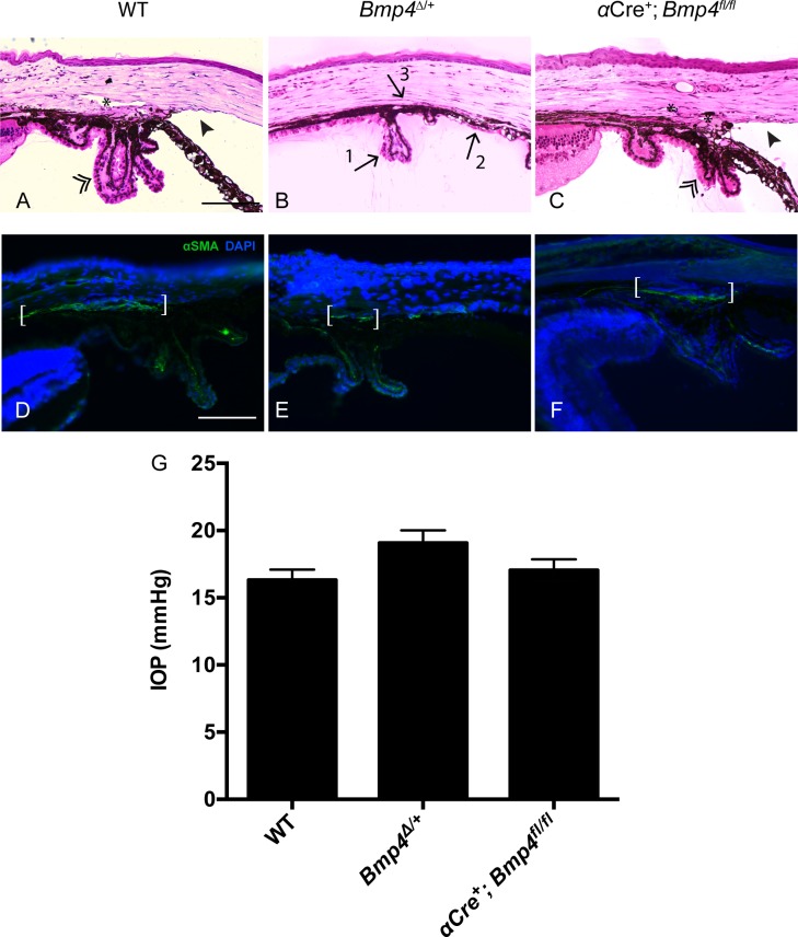
Fig 3. Tissues involved in IOP regulation do not require ciliary margin-derived BMP4.
Semi-thin plastic sections of Bmp4Δ/+ eyes stained with H&E show several morphological abnormalities including a hypoplastic ciliary body (arrow #1), trabecular meshwork (arrow #2), and iris, and an absence of Schlemm’s canal (arrow #3; B). Note normal morphology of ciliary body (double arrowhead), iridocorneal angle (solid arrowhead), and Schlemm’s canal (asterisk) in WT as well as αCre+; Bmp4fl/fl eyes (A, C; n = 3 per genotype). Note there is some variability in the appearance of ciliary processes and the extent of visible Schlemm’s canal in both WT and αCre+; Bmp4fl/fl mice, however both represent normal iridocorneal angles. Extensive expression of α-SMA is observed in the trabecular meshwork (white brackets) of WT and αCre+; Bmp4fl/fl mice (D, F), whereas a smaller area of immunostaining is noted in Bmp4Δ/+ mice (E; n = 3 per genotype). There was no significant difference between IOP measurements in adult mice between any of the three groups (G; WT n = 37, αCre+; Bmp4fl/fl n = 22m, p = 0.8237; WT n = 37, Bmp4Δ/+ n = 26; one-way ANOVA, p = 0.0502). Means and SEM are displayed. Scale bars in (A-F) represent 100μm.

