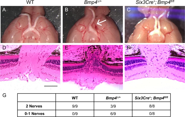
Fig 5. Central optic cup neuroepithelium-derived BMP4 is not required for optic nerve formation.
Control animals display normal gross optic nerve and optic chiasm morphology (A), as well proper axonal exit at the optic nerve head shown in plastic sections (D). Bmp4Δ/+ mice are often missing one (arrow) or both nerves (B), and retinal ganglion cell axons do not properly exit the eye (E). Optic nerves of Six3Cre+; Bmp4fl/fl mice appear indistinguishable from controls (C, F). The total number of brains examined for each strain is shown in the table below (G). At least 4 eyes per genotype were sectioned for histology. Scale bars in (D-F) represent 100μ.

