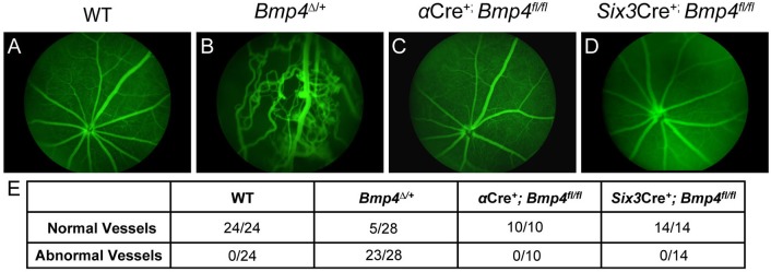
Fig 6. The retinal vasculature is properly organized following loss of optic cup neuroepithelium-derived BMP4.
Fundus exams using fluorescein angiography reveal highly abnormal vasculature patterning in Bmp4Δ/+ mice (B), however all αCre+; Bmp4fl/fl and Six3Cre+; Bmp4fl/fl mutants examined display normal vasculature formation (C, D). The total number of eyes examined for each strain is shown in the table below (E). Abnormal vessels were defined as blood leakage, irregularly arranged retinal capillaries, and vessel protrusion into the vitreous body.

