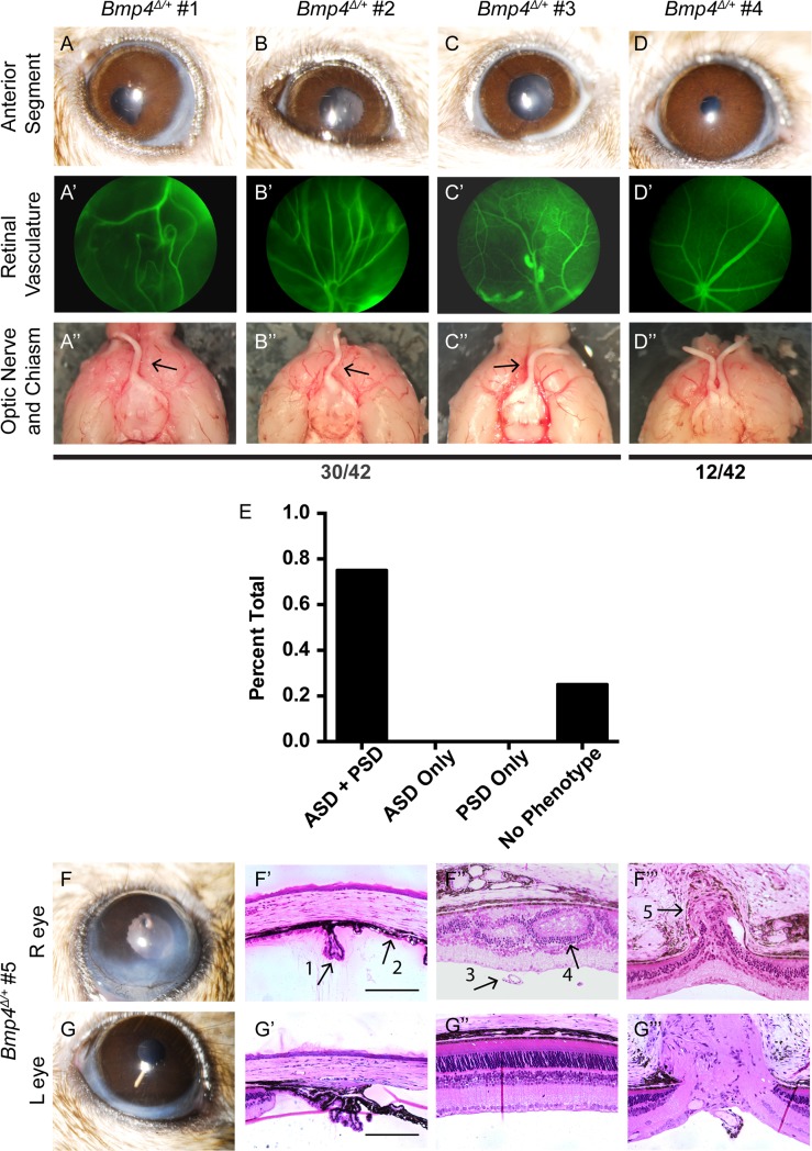
Fig 7. Anterior and posterior abnormalities occur concurrently or not at all.
Slit lamp images, fundus exams, and gross optic nerve morphology was assessed sequentially in 42 eyes of Bmp4Δ/+ mice. 30/42 eyes displayed each of the following: ASD, abnormal vasculature, and absence of an optic nerve (A-C, A’-C’, A”-C”). The remaining 12 eyes appeared normal in each category (D-D”). None of the eyes examined presented with only anterior segment dysgenesis or only posterior segment dysgenesis (E). An additional cohort (n = 6 eyes; 3 phenotypically abnormal and 3 with phenotypically normal based upon in vivo imaging) was assessed with plastic histology following slit lamp and fundus examination. Representative images from the same animal illustrate that the phenotypic presentation of the anterior segment (F, G) always predicts the presence or absence of morphological disruption in the posterior segment (F”, F”‘, G”, G”‘). Arrows represent: hypoplastic ciliary body (#1, F’), iridocorneal adhesion (#2, F’), persistent hyaloid vasculature (#3, F”), disrupted retinal lamination (#4, F”), and obstructed optic nerve exit (#5, F”‘). Scale bars represent 100μ.

