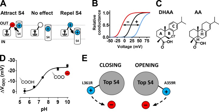
Figure 1.
The lipoelectric mechanism. (A) A compound binds with its hydrophobic anchor in the lipid membrane. The charged effector, attached to the anchor, electrostatically affects the positively charged voltage sensor (S4). (B) Compounds in A affect the voltage dependence of the channel opening. The direction of the shift depends on the valence of the charge. (C) Structures of DHAA and AA. The nomenclature of the ring structure and location of carbon 7 (C7) is shown in DHAA. (D) pH dependence for the effect of 100 µM DHAA on the 3R Shaker KV channel (updated from Ottosson et al., 2015). The carboxyl group is shown in either its protonated or deprotonated form. Error bars represent mean ± SEM (n = 4–8). (E) A negatively charged compound, located close to the voltage sensor S4, either facilitates or hinders channel opening by affecting the rotation of the voltage sensor S4. The direction depends on which side of S4 an arginine (R) is located.

