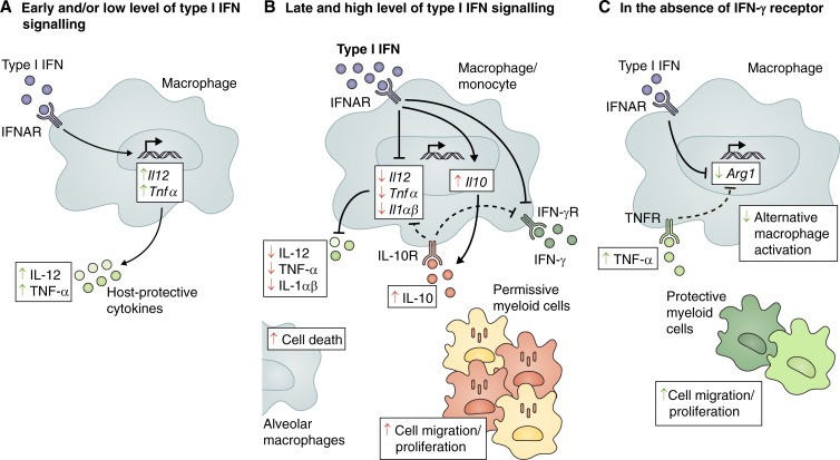
Figure 2.
Foe- and friendly-like effects of type I IFN during M. tuberculosis infection. Type I IFN has been reported to play both negative (red arrows) and positive (green arrows) functions during M. tuberculosis infection. (A) Tonic levels of autocrine type I IFN signaling prime the production of protective cytokines IL-12 and TNF-α. (B) However, high and sustained levels of type I IFN promote the production of IL-10 and inhibit the production of protective cytokines IL-12, TNF-α, IL-1α, and IL-1β. IL-10 mediates a suppressive feedback loop, contributing to the decreased production of IL-12 and TNF-α. Type I IFN also inhibits myeloid cell responsiveness to IFN-γ by both IL-10–dependent and independent mechanisms, suppressing IFN-γ–dependent host-protective immune responses. In addition, type I IFN can promote cell death in alveolar macrophages and accumulation of permissive myeloid cells at the site of infection. (C) In the absence of the IFN-γ receptor, type I IFN inhibits Arg1 expression directly or indirectly by increasing TNF-α levels, thus regulating macrophage activation toward a more protective phenotype. Type I IFN signaling can also promote the recruitment, differentiation, and/or survival of protective myeloid cells that control pathology at the site of infection. Arg1, arginase 1; IFNγR, IFN-γ receptor; IL-10R, IL-10 receptor; TNFR, TNF-α receptor.

