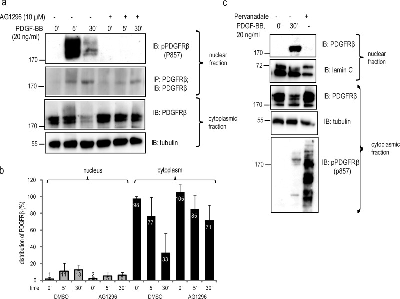
Figure 2.
PDGF-BB–induced nuclear transport of PDGFRβ is not quenched by receptor kinase inhibition. (a) PDGFRβ translocation to the nucleus upon inhibition of PDGFRβ kinase activity. Nuclear extracts were immunoblotted for pTyr857-PDGFRβ (top) or immunoprecipitated and immunoblotted with the PDGFRβ antibody ctβ (second panel). Cytoplasmic extracts were immunoblotted for PDGFRβ with the ctβ antibody (third panel) or α-tubulin (bottom). (b) Quantification of PDGFRβ distribution upon inhibition of receptor tyrosine kinase activity with AG1296. Immunoblots were quantified as described in Fig. 1. Error bars indicate SD. Quantification was based on four independent experiments. (c) Cells were stimulated with PDGF-BB or phosphatase activity was inhibited with sodium pervanadate, and lysates were immunoblotted with total PDGFR antibody (Y92) or lamin A/C for nuclear fractions (top two panels) or total PDGFRβ, α-tubulin, and pPDGFRβ antibody for cytoplasmic fractions (three bottom panels). Molecular mass was measured in kilodaltons. IB, immunoblotting; IP, immunoprecipitation.

