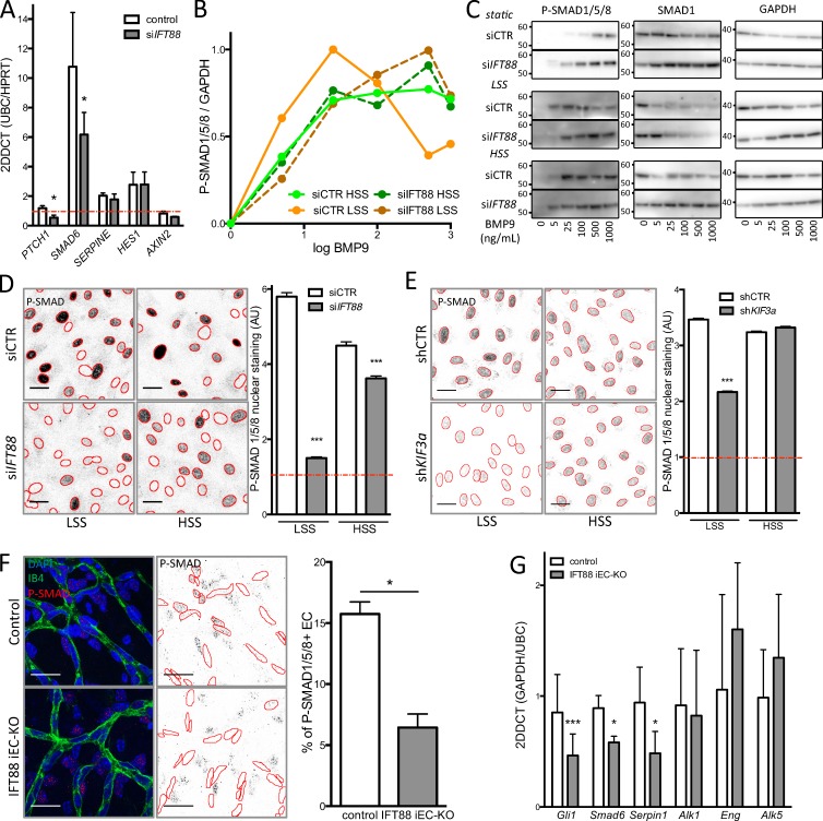
Figure 4.
Loss of the primary cilium leads to increased migration and blocks the differential response to BMP9 under flow. (A) quantitative PCR was used to assess levels of SHH (PTCH1), BMP (SMAD6), TGFβ (SERPINE), Notch (HES1), and Wnt (AXIN2) signaling in ECs transfected with siIFT88 or siCTR under LSS conditions (n = 5; all conditions were normalized to 1 for siCTR under the static condition [red line, n = 4]; mean± SEM; two-sided Wilcoxon test; *, P < 0.05). (B) Dose–response curves were generated for p-SMAD1/5/8 levels after BMP9 stimulation based on Western blot analysis (n = 3; mean: LSS = 2 dyn/cm2; HSS = 20 dyn/cm2). (C) Representative images of the Western blots used for the dose–response analysis. Molecular masses are indicated on the images based on prestained protein standard. (D) Representative images (red outline based on DAPI segmentation) and quantification of p-SMAD1/5/8 nuclear staining (PFA fixation) in ECs transfected with siCTR or siIFT88 and treated with 10 pg/ml BMP9 (the red line represents nontreated cell level of staining; n = 3; between 1,900 and 3,300 cells per condition; mean± SEM [of cell individual values]; two-sided Mann–Whitney U test; ***, P < 0.001). Bars, 30 µm. (E) Representative images and quantification of p-SMAD1/5/8 nuclear staining (PFA fixation) in ECs transfected with shCTR or shKIF3a and treated with 10 pg/ml BMP9 (the red line represents nontreated cell level of staining; n = 3; between 1,900 and 3,300 cells per condition; mean± SEM [of cell individual values]; two-sided Mann–Whitney U test; ***, P < 0.001). Bars, 30 µm. (F) Representative images of immunofluorescent staining of p-SMAD1/5/8 in the retina of IFT88 iEC-KO mice or littermate control and quantification of the nuclear signal in ECs. Positive staining in surrounding cells is still visible in the IFT88 iEC-KO retina (control, n = 5; IFT88 iEC-KO, n = 7, blue: DAPI; red: p-SMAD1/5/8; green: Isolectin; mean± SEM; two-sided Wilcoxon test; *, P < 0.05). Bars, 20 µm. (G) qPCR analysis was performed on ECs extracted from IFT88 iEC-KO (gray) and littermate retinas (white; n = 7; mean± SEM; two-sided Wilcoxon test; *, P < 0.05; ***, P < 0.001).

