Figure 1.
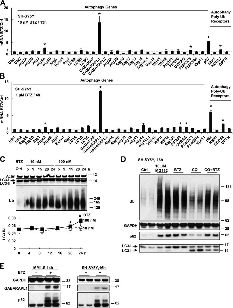
Proteasome inhibitor treatment causes rapid induction of p62 and GABARAPL1 but not other Atg genes or Ub receptors. (A and B) SH-SY5Y cells were treated with 10 nM BTZ for 13 h (A) or 1 µM for 4 h (B). The mRNAs of all Atg proteins or Ub receptors were measured in this and other figures by real time RT-PCR. (C) Autophagy was activated only after prolonged treatment with a high concentration of proteasome inhibitor. SH-SY5Y cells were treated with 10 or 100 nM BTZ for 5–24 h. To measure autophagy, the levels of LC3-I and lipidated (autophagosome-bound) LC3-II were measured (upper) by Western blotting, and the LC3-II/I ratio was quantified (lower). *, LC3-II/I ratio in cells treated with 100 nM BTZ is higher than that in untreated cells (P < 0.05; n = 2). (D) Treating SH-SY5Y cells for 16 h with 50 µM chloroquine (CQ) prevented the degradation of LC3-II, but cells treated with BTZ (10 nM) and CQ did not accumulate more LC3-II than cells treated only with CQ. (E) After SH-SY5Y or MM1.S cells were treated with 10 nM BTZ, levels of p62 and GABARAPL1 proteins were greatly increased. *, P < 0.05. Molecular masses are given in kilodaltons. Error bars indicate SD.

