Figure 3.
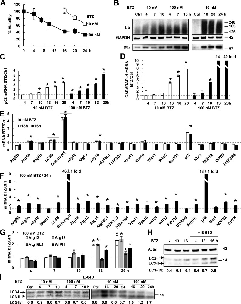
Upon BTZ treatment, RPMI 8226 cells rapidly induce p62 and GABARAPL1, but only after prolonged treatment do they induce other Atg genes and activate autophagy. (A) The effect of BTZ treatment on the viability of RPMI 8226 cells was measured by MTS assay. n = 3. (B) BTZ treatment (especially with high concentrations) caused Ub conjugate buildup within 4–7 h and increased p62 protein levels. (C and D) Upon BTZ treatment, cells rapidly induced mRNAs for p62 (C) and GABARAPL1 (D). (E) Cells induced mRNAs for p62 and GABARAPL1, but not most other Atg genes when treated with 10 nM BTZ for 13 or 16 h. (F) Cells treated with 100 nM BTZ for 24 h induced almost all Atg genes. (G) Cells treated with 100 nM BTZ induced most Atg genes only at 16 or 20 h. (H) Treating control or BTZ-treated cells with 10 µM E-64D for 1 h to inhibit lysosomal proteases increased the LC3-II level and the LC3-II/I ratio. (I) The LC3-II/I ratio was measured after E-64D treatment as in H to evaluate autophagic activity. For this ratio to rise twofold, cells need to be treated with a high concentration (100 nM) of BTZ for ≥20 h. *, P < 0.05. Molecular masses are given in kilodaltons. Error bars indicate SD.

